. 2022 Sep 9;42(7):1925–1939. doi: 10.1148/rg.220073

Figure 6.

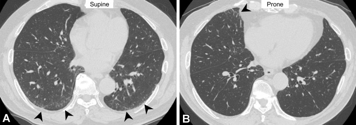
Dependent abnormality in a 72-year-old man with rheumatoid arthritis. (A) Axial CT image obtained with the patient supine shows ground-glass abnormality (arrowheads) in the subpleural lung area. (B) On the axial CT image obtained with the patient prone, the ground-glass abnormality has disappeared in the subpleural area but is seen in the middle lobe of the right lung (arrowhead). These findings are considered to indicate transient lung atelectasis.

Dependent abnormality in a 72-year-old man with rheumatoid arthritis. (A) Axial CT image obtained with the patient supine shows ground-glass abnormality (arrowheads) in the subpleural lung area. (B) On the axial CT image obtained with the patient prone, the ground-glass abnormality has disappeared in the subpleural area but is seen in the middle lobe of the right lung (arrowhead). These findings are considered to indicate transient lung atelectasis.