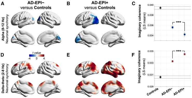
Figure 2.
Distinctive regional patterns of neuronal synchrony deficits in Alzheimer’s disease patients with and without epileptiform activity. (A–F) Statistical comparisons of imaginary coherence between groups. Within the alpha band, AD-EPI− and AD-EPI+ showed significantly reduced imaginary coherence compared to controls, over bilateral occipital cortices, with more extensive regional involvement in AD-EPI+ (A and B). Pairwise comparison showed AD-EPI+ with significantly lower alpha imaginary coherence than AD-EPI− (C). AD-EPI− and AD-EPI+ showed significantly increased delta–theta imaginary coherence compared to controls, over bilateral frontal and parietal cortices, with more extensive involvement in AD-EPI+ (D and E) and pairwise comparison showed AD-EPI+ with significantly higher delta–theta imaginary coherence than AD-EPI− (F). Each brain rendering depicts the t-maps from voxelwise comparison of global imaginary coherence between groups. The colour maps are thresholded with a cluster correction of 30 voxels (P < 0.01) and at 5% FDR. (C and F) depict least squares (LS)-means and 95% confidence-limits (n = 30, AD-EPI+; n = 20 AD-EPI−; n = 35 age-matched controls). Alzheimer’s disease = Alzheimer’s disease; AD-EPI− = Alzheimer’s disease patients without epileptiform activity; AD-EPI+ = Alzheimer’s disease patients with epileptiform activity; IC = imaginary coherence.

