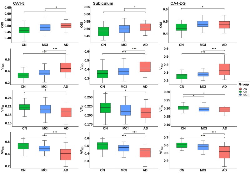
Figure 2.
Group differences of Cortical-NODDI-derived ODI, VISO, VFIC and VFEC in the hippocampal subfields among the cognitively normal, MCI and Alzheimer’s disease participants. The comparison was conducted using general linear model with age, sex, level of education, APOE ε4 status and total intracranial volume as covariates. Multiple comparisons across three regions of interest (i.e. subfields) were adjusted by FDR using the Benjamini-Hochberg criterion (α = 0.05). *PFDR < 0.05; **PFDR < 0.01; ***PFDR < 0.001. AD = Alzheimer disease; CN = cognitively normal.

