Figure 1.
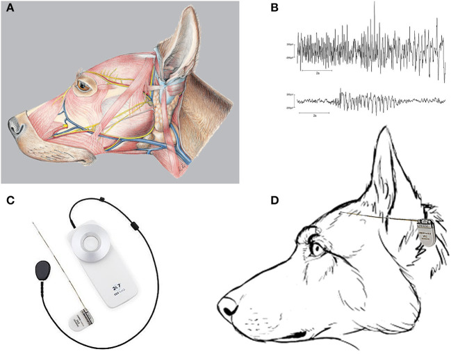
The musculature of the dog's head and seizure recording by a subscalp EEG device. (A) Illustrates the massive musculature of the dog's head (see also Figure 2D), which produces artifacts when applying scalp EEG electrodes. The picture was taken from Reese et al. (15). (B) Two different EEG seizures were recorded from Beagle dogs with subscalp electrodes inserted below the temporalis muscle on the skull to minimize electromyographic artifacts [from Authier et al. (16)]. EEG data were obtained using telemetry transmitter leads with bipolar derivations according to the internationally standardized 10–20 system, using Cz-Oz derivations as previously described (17). (C) The 24/7 EEG™ SubQ system from UNEEG Medical (Lillerød, Denmark), involving a small ceramic implant that consists of an electrode house containing an inductive coil for the transfer of power and data and a wire with three leads (electrodes). The center electrode is used for reference, and the recordings, therefore, have two bipolar channels. An external device supplies the implant with power, receives and stores the measured EEG signals, and when coupled with a smartphone can wirelessly stream the recorded EEG data automatically to a cloud environment. (D) Schematic illustration of how the 24/7 EEG™ SubQ system may fit to the size of a dog's head. The device will be tunneled under the temporalis muscle and proximate to the dog's skull (not illustrated; see text).

