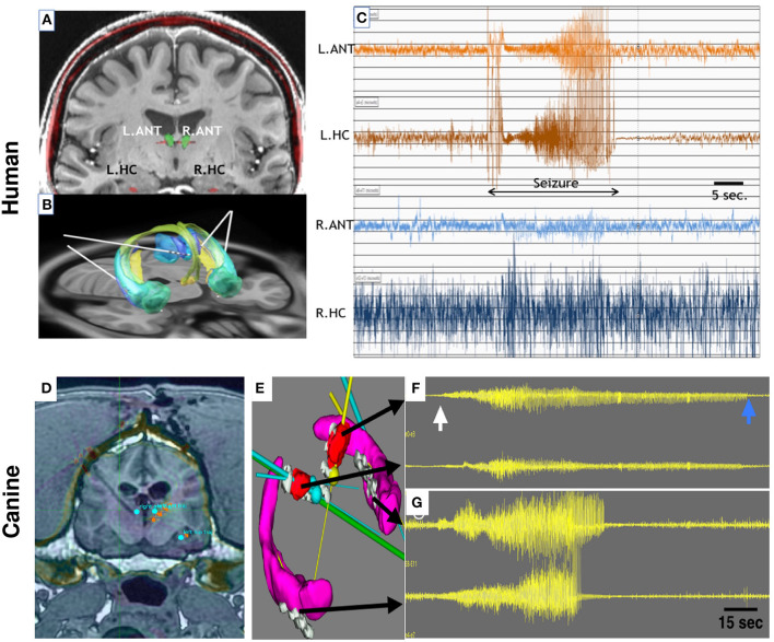
Figure 2.
Bilateral Anterior Nucleus Thalamus (ANT) and Hippocampus (HPC) targets for mesial temporal lobe epilepsy in human (A–C) and canine (D–G) epilepsy. (A) 3T MRI of a human brain with targets for ANT(green) and HPC (red) electrodes labeled. (B) Papez circuit with ANT (blue) and HPC—AMG (green) targets and implanted electrode leads (white lines). (C) Spontaneous focal seizure with simultaneous left ANT and HPC involvement. Clinically the patient had intense fear and loss of awareness. The seizure has synchronous left ANT and HPC onset without the evolvement of contralateral ANT or HPC. (D) 3T MRI and stereotactic targeting in the canine brain. The bilateral ANT and HPC targets are highlighted. The coronal MRI shows the massive cranial musculature surrounding the canine skull. (E) Bilateral HPC (purple) and ANT (red) target volumes and implanted electrodes (gray). The multiple color straight lines indicate target trajectories for electrode leads. (F,G) Four channels of intracranial EEG. From top to bottom: Left ANT, Right ANT, Left HPC, and Right HPC recordings. The onset of a seizure (white arrow) and the seizure offset (blue arrow) are clearly evident in both HPC and ANT recordings, but interestingly the seizure terminates in all electrodes at different times. The longest seizure discharge is in the left ANT (top trace). See text for details. ANT, anterior nucleus of the thalamus; HPC, hippocampus. Adapted from Sladky et al. (18).

