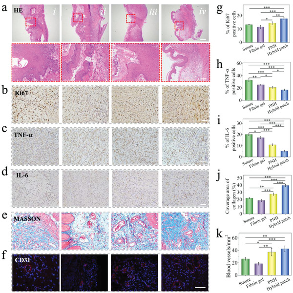
Figure 6.

The in vivo evaluation of the hybrid patch on a rat gastric perforation model. Representative a) HE staining, b–d) Ki67, TNF‐α, and IL‐6 immunohistochemistry staining, e) Masson staining, and f) CD 31 immunofluorescence staining images of the wound area after treating with i) suture, ii) fibrin gel, iii) PNH, and iv) hybrid patch for 7 days, respectively. The scale bar in (a) is 500 µm, in the enlarged images is 100 µm. The scale bar in (b)‐(f) is 50 µm. g–i) Quantification of Ki67, TNF‐α, and IL‐6 positive cells. j) Quantification of collagen coverage area. k) Quantification of CD31 labeled blood vessels. n = 3, biologically independent samples. Data are showed as mean ± SD. *p < 0.05; **p < 0.01; ***p < 0.001, using one‐way ANOVA followed by post‐hoc test.
