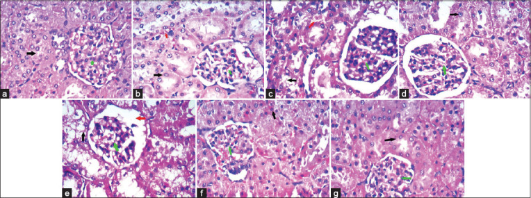
Figure-3.

(a) Cloudy swelling of the renal tubular epithelial cells and over condensed glomerular tuft and capsule of control rabbits, glomeruli showed capillary tuft (green arrow), thin Bowman’s space, and mesangial cells, while tubules lined by columnar cells (black arrow) with eosinophilic cytoplasm while interstitium showed blood vessels and stroma. (b) Kidney of rabbits treated with 20 mg salinomycin/kg ration showed dilatation of Bowman’s capsule, star-shaped lumen of BCT, hydropic degeneration (black arrow) of tubular epithelial cells, glomeruli (green arrow) showed a slight increase in mesangial cellularity and stroma congestion (red arrowhead). (c) Kidney of rabbits treated with 40 mg salinomycin/kg ration showed marked glomerular atrophy (green arrow) and hydropic degeneration (black arrow) of tubular epithelial cells with dilatation of Bowman’s space (red arrowhead) with increased mesangial cellularity. (d) Kidney of rabbits treated with 20 mg salinomycin/kg ration + 6.5 mg silymarin/kg BW showed improvement evident as only residual minimal stromal congestion (red arrowhead) and glomerular congestion (green arrow) with regular tubules (black arrow). (e) Kidney of rabbits treated with 40 mg salinomycin/kg ration + 13 mg silymarin/kg BW showed marked improvement evident as regular tubules (black arrow) and glomeruli (green arrow). (f) Kidney of rabbits treated with 6.5 mg silymarin/kg BW formed of glomeruli and tubules, glomeruli showed capillary tuft (green arrow), thin Bowman’s space, and mesangial cells, while tubules lined by columnar cells (black arrow) with eosinophilic cytoplasm while interstitium showed blood vessels and stroma. (g) Kidney of rabbits treated with 13 mg silymarin/kg BW formed of glomeruli and tubules, glomeruli showed capillary tuft (green arrow), thin Bowman’s space, and mesangial cells, while tubules lined by columnar cells (black arrow) with eosinophilic cytoplasm while interstitium showed blood vessels and stroma (H&E stain. 400×).
