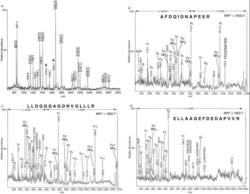
FIG. 3.
Analysis of EF-Tu homolog (spot 4) by mass spectrometry. (A) MALDI-TOF MS peptide mass fingerprint spectrum produced by in-gel tryptic digestion of gel spot 4, pooled from four 2-D gels. (B through D) MALDI-PSD mass spectrum of a tryptic peptide with (B) MH+ at m/z of 1,405.4, (C) MH+ at m/z of 1,682.7, and (D) MH+ at m/z of 1,802.7. Fragment ions detected in the PSD analysis were used to search the protein databases with MS-Tag. All three peptides were identified as belonging to EF-Tu proteins from both M. tuberculosis and M. leprae. In the mass fingerprint spectrum (panel A) the peak at m/z of 857.4 was identified as SQRYFR from MALDI-PSD data and was identified as the sole peptide in spot 4 belonging to a minor contaminating protein (NCBI accession no. 1722931) of unknown function. See legend to Fig. 2 for an explanation of peak labels.

