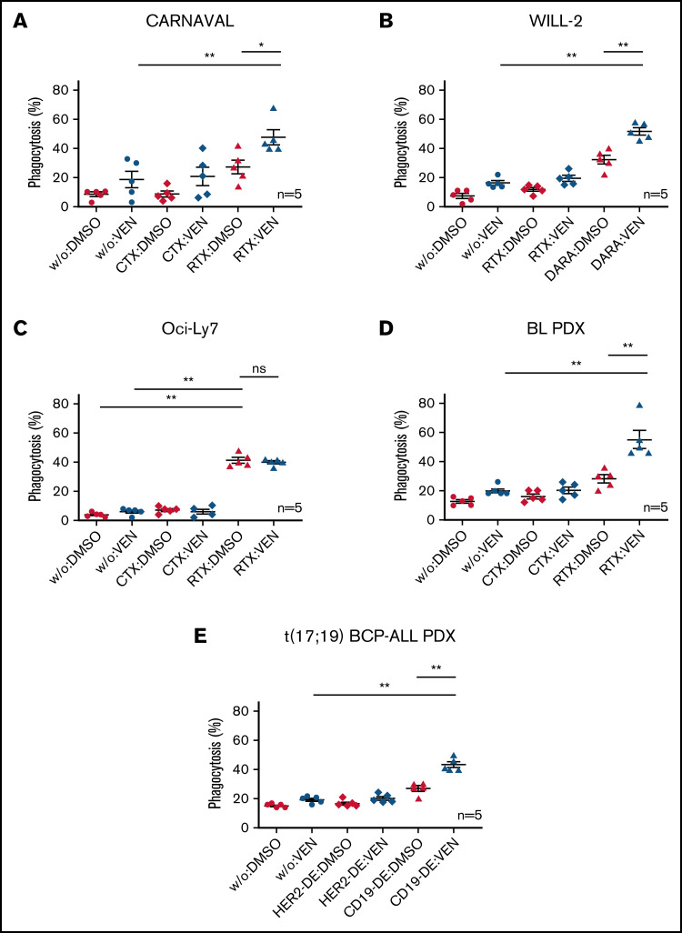
Figure 2.
ADCP in Bcl-2–expressing DHL cell lines and PDX samples on combination of VEN and therapeutic antibodies. Percentages of cells phagocytosed by human macrophages with VEN/antibody combinations compared with VEN or antibody (RTX, DARA, CD19-DE) alone. (A) CARNAVAL cells after treatment with 1 nM VEN for 12 hours, RTX, and the control antibody CTX. (B) WILL-2 cells treated with 1 nM VEN for 12 hours, DARA, and the control antibody RTX. (C) Oci-Ly7 cells after treatment with 1 nM VEN for 12 hours, RTX, and the control antibody CTX. (D) ADCP in BL PDX sample subjected to 1 nM VEN for 12 hours, RTX, and the control antibody CTX. (E) t(17;19)-positive BCP-ALL PDX sample treated with 1 nM VEN for 12 hours, CD19-DE, and the control antibody HER2-DE (a version of trastuzumab containing the same modification in the Fc part of the antibody as CD19-DE). DMSO, solvent control; w/o, no antibody. Phagocytosis was determined as the percentage of macrophages with completely ingested carboxyfluorescein succinimidyl ester green–positive cells by counting in total 100 macrophages by at least 3 independent observers. Each dot represents an independent experiment with different human donors. Data are presented as mean ± standard error of the mean (SEM) from independent experiments with 5 healthy blood donors. Statistical analysis: ns, not significant; *P < .05; **P < .005; Mann-Whitney test. All antibodies used in vitro were applied to a final concentration of 10 µg/mL.

