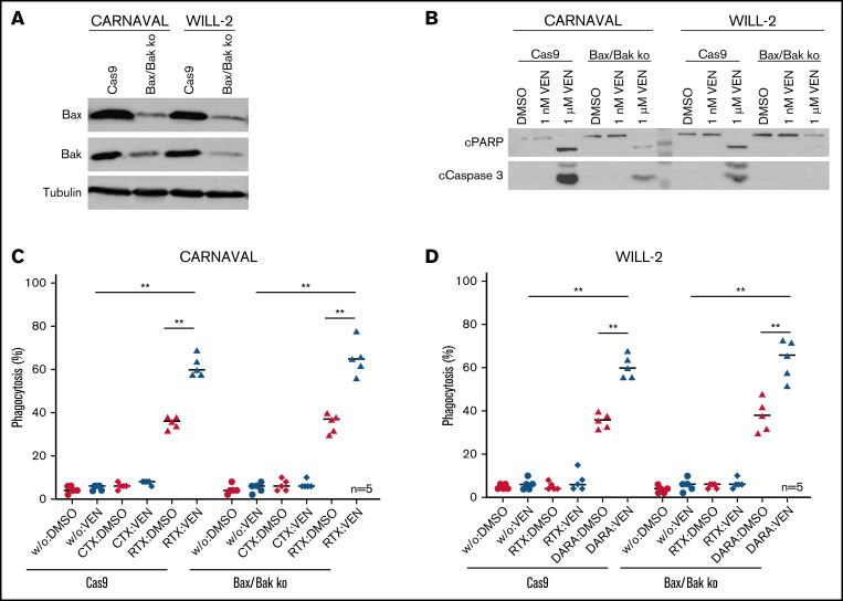
Figure 5.
VEN-mediated phagocytosis in Bax/Bak-deficient DHL cell lines. (A) Protein levels of Bax and Bak in CARNAVAL and WILL-2 Bax/Bak ko cells and control cells (Cas9) analyzed by Western blot. (B) Examination of cPARP and cCaspase 3 in CARNAVAL and WILL-2 Bax/Bak ko cells control cells (Cas9) subjected to 1 nM or 1 µM VEN for 12 hours. (C) ADCP in CARNAVAL deficient for Bax/Bak or control cells (Cas9) after treatment with 1 nM VEN for 12 hours, RTX, and the control antibody CTX. (D) ADCP in WILL-2 lacking Bax/Bak or control cells (Cas9) treated with 1 nM VEN for 12 hours, DARA, and the control antibody RTX. Data are presented as mean ± SEM from independent experiments with 5 healthy blood donors. Statistical analysis: *P < .05; **P < .005; Mann-Whitney test. All antibodies used in vitro were applied to a final concentration of 10 µg/mL. Tubulin served as a loading control.

