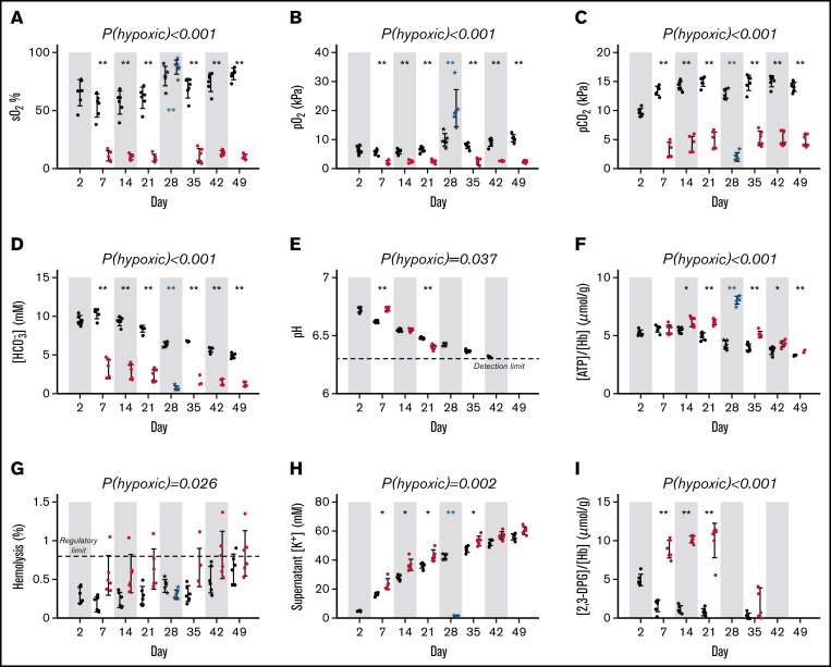
Figure 3.
Standard quality measurements of units. Red cell concentrates (RCC) upon pooling or when subsequently split and stored under the hypoxic storage system (red dots), or standard normoxic storage (black dots) and rejuvenated (blue dots). (A) Oxygen saturation (sO2; %). (B) Partial pressure of O2 or (C) carbon dioxide (pCO2; kPa). (D) bicarbonate concentration ([HCO3−] mmol/L). (E) pH; note the lower detection limit is 6.3. (F) Cellular (ATP) (μmol/gHb) measured by assay. (G) Degree of hemolysis. (H) Supernatant potassium concentration (mmol/L). (I) [2,3-DPG] (μmol/gHb) measured by assay (measured for storage days 2, 7, 14, 21, and 35 only). Error bars represent mean ± standard deviation (n = 6). Significance values for statistical tests were denoted *P < .05, **P < .01 for hypoxic vs standard storage (repeated measures 2-way ANOVA, followed by multiple comparisons test) and for pre- and postrejuvenation (paired 2-sample t-test).

