
FIG. 1.
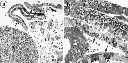
Epithelial sheets detach from rabbit appendix after treatment with EDTA. (a) Light micrograph of rabbit appendix tissue after treatment with EDTA for 2 h. Epithelial sheets are being released from the lamina propria (LP). M cells (arrows) are recognizable by their pockets filled with lymphocytes. The lymphocytes in the follicle are retained in the mucosal tissue (LF). (b) Light micrograph of a sectioned pellet of isolated epithelial sheets. The cells are still attached to form monolayer sheets containing M cells (arrows). Some layers from the villus epithelium display goblet cells (arrowheads).