
Fig. 6.
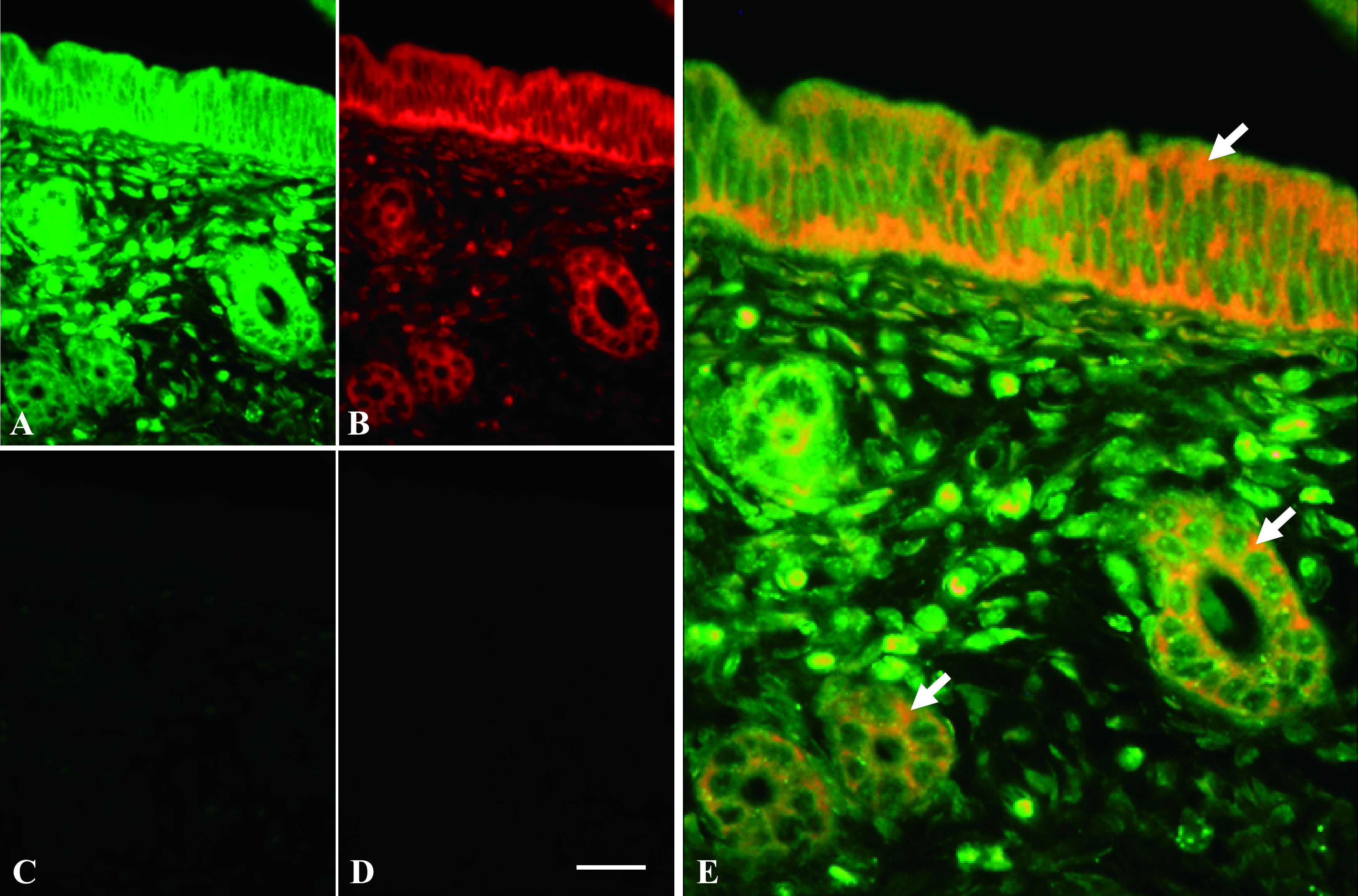
ERα mRNA and 28S rRNA expression in mouse uterine tissue using ISH. Uterine tissue was hybridized with complementary MB probe for 28S rRNA (green) (A) and antisense MB probe for ERα mRNA (red) (B). Both images were merged in (E), and arrows indicate co-localization of ERα mRNA and 28S rRNA expressions. Adjacent uterine section was hybridized with homologous MB probe for 28S rRNA (C) and sense MB probe for ERα mRNA (D). Magnification ×400. Bar = 20 μm.