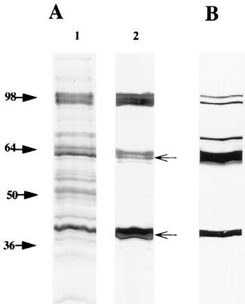
FIG. 1.

(A) Silver-stained SDS–10% polyacrylamide gel of C. pneumoniae EB (lane 1) and OMC (lane 2). The samples were boiled in SDS sample buffer. (B) Immunoblotting of C. pneumoniae EBs reacted with the polyclonal rabbit antiserum to C. pneumoniae OMC. The arrows indicate the migration of Omp2 (60 to 62 kDa) and MOMP (39.5 kDa).
