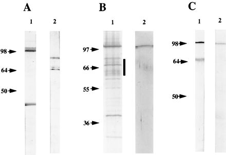
FIG. 7.

Analysis of the migration pattern of C. pneumoniae OMC proteins in SDS-PAGE. (A) C. pneumoniae OMC proteins were solubilized in SDS sample buffer. Then boiled (lane 1) and unboiled (lane 2) proteins were separated by SDS-PAGE and subjected to immunoblotting with MAb 24.1.44. (B) Silver staining of SDS-PAGE-separated C. pneumoniae OMC proteins solubilized in SDS sample buffer without boiling (lane 1). The bands marked with a vertical bar were excised from a Cu2+-stained gel, boiled in SDS sample buffer, rerun in SDS-PAGE, and silver stained (lane 2). (C) Alternatively, the bands marked with the bar in panel B were excised, boiled and subjected to immunoblotting with PAbOmp4 (lane 1) and MAb 24.1.44 (lane 2).
