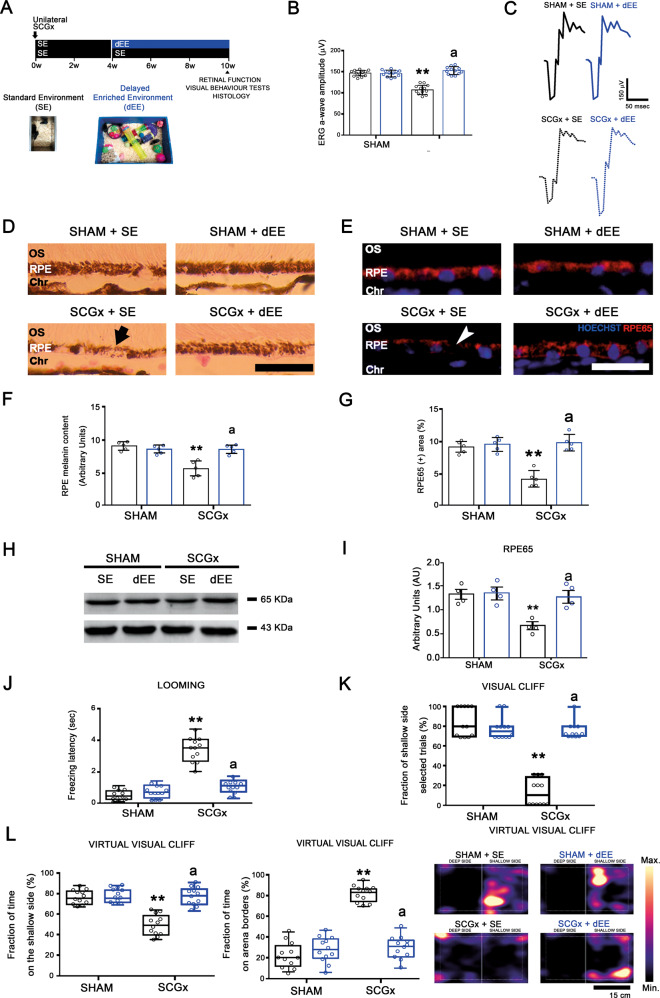
Fig. 5. Effect of the delayed EE exposure (dEE) on the retinal function and temporal RPE structural alterations and visual behaviour tests at 10 weeks post-SCGx.
A Experimental protocols. B EE starting at 4 weeks post-SCGx completely reversed the decrease in the ERG a-wave amplitude (representative traces shown in C). Data are mean ± SEM (n: 12 animals per group), **P < 0.01 vs. SE animals with sham-treated eyes; a: P < 0.01 vs. SE animals with SCGx-treated eyes, by Tukey’s test. D–I SCGx induced a decrease in the melanin content (arrow) and RPE65-immunoreactivity (arrowhead) and protein levels at the temporal RPE in SE animals. The delayed exposure to EE totally reversed these alterations. Shown are representative photomicrographs from 5 eyes/group at 800 μm temporally from the ONH. OS PR outer segments, RPE retinal pigment epithelium, Chr choroid. Scale bars = 25 μm. Data are mean ± SEM (n: 5 eyes per group), **P < 0.01 vs. sham-treated eyes from SE animals; a: P < 0.01 vs. SCGx-treated eyes from SE animals, by Tukey’s test. Data are mean ± SEM (n: 5 homogenates per group), **P < 0.01 vs. sham-treated eyes from SE animals, by Tukey’s test. J–L The delayed exposure to EE reversed the worse performance in the looming, visual cliff and virtual visual cliff tests induced by SCGx in SE-housed animals. **P < 0.01 vs. SE animals with sham-treated eyes; a: P < 0.01 vs. SE animals with SCGx-treated eyes, by Tukey’s test (n: 12 animals per group).

