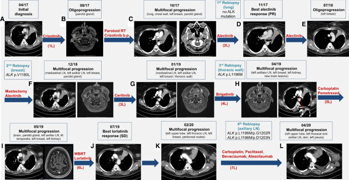
Figure 1.
Radiologic findings over the course of the disease. (A) Chest computed tomography (CT) scan at initial diagnosis. (B) Head magnetic resonance imaging (MRI) 4 mo after crizotinib start with oligoprogression in the right parotid gland. (C) Chest and cranial CT scans with multifocal progression after radiotherapy. (D) Chest CT scan showing response to alectinib. (E) Chest CT scan showing oligoprogression in the left breast 8 mo later. (F) Chest CT and head MRI showing multifocal progression after partial mastectomy. (G,H) Chest CT and head MRI showing multifocal progression under certinib and brigatinib. (I) Chest CT and head MRI showing multifocal progression under carboplatin-pemetrexed. (J) Chest CT showing response to lorlatinib. (K) Chest CT and head MRI showing multifocal progression under lorlatinib. (L) Chest CT showing multifocal progression under carboplatin-paclitaxel-bevacizumab-atezolizumab; the patient died 3 wk later. (L) Treatment line, (RT) radiotherapy, (PR) partial response, (SD) stable disease, (LN) lymph nodes.

