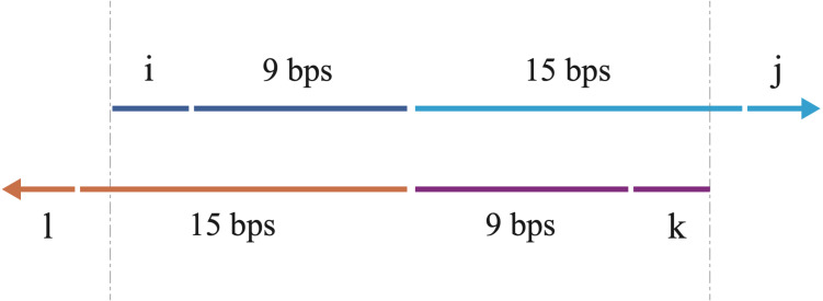
Figure 2. A schematic illustration of the overlapping primers.
The arrows show the 5′–3′ direction of DNA. The primers are partially complemented with 3′ overhangs. The upper arrow indicates the forward overlapping primer, consisting of the nucleotides (blue) from the 3′ end of the sense strand of the first DNA fragment and the nucleotides (cyan) from the 5′ end of the sense strand of the second DNA fragment. The lower arrow indicates the reverse overlapping primer, consisting of the nucleotides (purple) from the 3′ end of the antisense strand of the second DNA fragment and the nucleotides (orange) from the 5′ end of the antisense strand of the first DNA fragment. The length of each part is labeled, with i, j, k, l all ranging from 0 to 3 bps (3 bps included). The variations of i, j, k, l enable us to walk through all possible combinations for the overlapping primers. The vertical gray dashes show the overlapping region between the two primers, which is also used for bridging during overlap extension PCR to acquire the full-length chimera DNA sequence.

