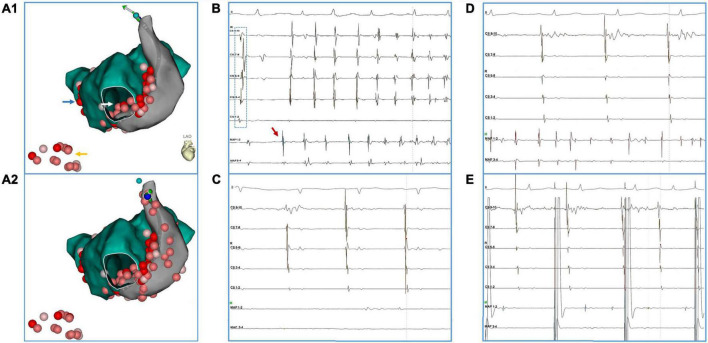
FIGURE 1.
A representative case of PLSVC isolation. The patient (#16) had AF recurrence after a previous ablation procedure in which only PVI was performed. (A1,A2) Blue arrow: touch up ablation at RPV. Yellow arrow: linear ablation at CTI. White arrow: CFAE at inferior LA. (B) Ectopy (red arrow) from distal PLSVC triggered an episode of AF. Notably, during the sinus beat, activation at the CS catheter presented a bracket-like sequence, which was probably caused by an earlier breakthrough at mid-PLSVC by LA-PLSVC connections. (C) After ablation at LA-PLSVC connections [Visitag points in PLSVC in (A1)], AF terminated and sinus rhythm was restored. However, a mapping catheter positioned at the distal PLSVC could still record fibrillatory activities. (D) Further ablation at distal PLSVC (beyond the level of left superior PV) and ablation at the CS-PLSVC connections was conducted [Visitag points in PLSVC in (A2)] which resulted in the elimination of local potential and loss of capture of LA during pacing from distal PLSVC (E). (Abbreviations same as those in the main body).

