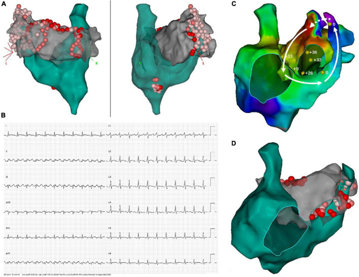
FIGURE 3.
Demonstration of twice ablation procedure in a patient with persistent AF and PLSVC. (A) In the first ablation procedure, PVI, as well as linear ablation at the LA roofline, MI line, and CTI line was performed. Extensive ablation targeting LA-PLSVC and CS-PLSVC connection, as well as high-frequency signals inside PLSVC, resulted in PLSVC isolation. (B) ECG of recurrent atrial flutter (AFL). (C) Bi-atrial AFL involving a connection between PLSVC and left atrial appendage. Sites where entrainment mapping was conducted were indicated by yellow dots, with the numerical value of post-pacing interval minus tachycardia cycle length (PPI-TCL) labeled by white numbers. The dotted arrow indicated conduction through epicardial connections between the left atrial appendage (LAA) and PLSVC, while the solid lines with arrowhead indicated conduction pathway through LAA, the anterior wall of LA, interatrial septum, septal aspect of the right atrium, coronary sinus and PLSVC. (D) Repeated ablation targeting resumed LA-PLSVC connections, as well as touch-up ablation at the roofline, was conducted in the redo-procedure.

