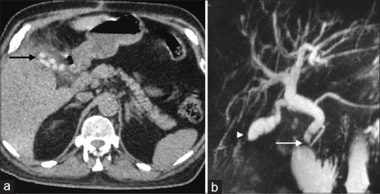
Figure 1.

(a) Computed tomography of the abdomen showing radio dense calculi in the residual gallbladder (arrow); (b) Magnetic resonance cholangiopancreatography showing a filling defect in the lower end of the dilated common bile duct (arrow) and a residual gallbladder (arrowhead)
