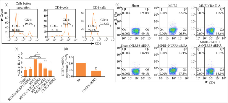
Figure 5. Inhibition of NLRP3 suppressed Th17 differentiation of CD4+ T cells. (a) Percentages of CD4+ T cells that were isolated by positive selection with MACS CD4 microbeads as shown by FCM. (b) The percentage of Th17 cells wasassessed by FACS. (b) Data were expressed as representative FACS images or (c) corresponding statistical graph (n ≥ 3).(d) The mRNA expression of NLRP3 in CD4+ T cells transfected with NLRP3 siRNA or their controls (n ≥ 3).
NLRP3: pyrin domain containing 3; MI/RI: myocardial ischemia-reperfusion injury; *P < 0.05; **P < 0.01; # P < 0.05 vs. normal control; IL: interleukin.

