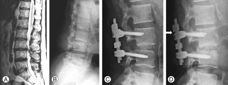
Fig. 5.

Representative case presentation. (A) Magnetic resonance imaging showing canal stenosis at L3/4. (B) Preoperative lateral radiograph showing degenerative spondylolisthesis at L3/4. (C) Postoperative lateral radiograph showing dynamization–posterior lumbar interbody fusion at L3/4. (D) Postoperative lateral radiograph after 2 years showing bone union with maintained local lordosis. A gap appeared between the stopper and connector (arrow).
