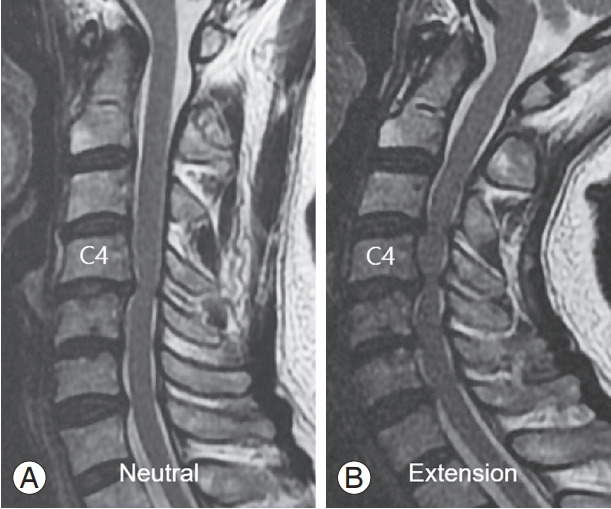
Fig. 2.

A 63-year-old female patient with degenerative cervical myelopathy. (A) Neutral sagittal magnetic resonance (MR) imaging of the cervical spine showed a C4–5 canal stenosis with signal change of the spinal cord. (B) Extension sagittal MR of the cervical spine showed more aggravated cord compression of C4–5 segment, as well as the cranial and caudal extension of the cervical canal stenosis to C3–7 levels.
