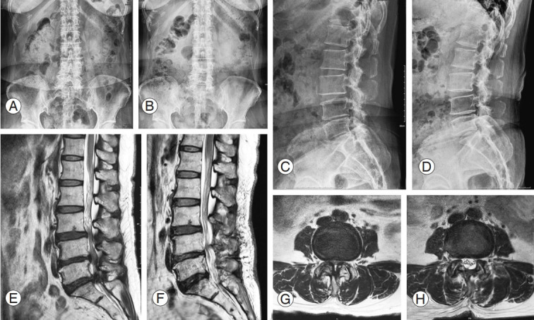
Fig. 1.

Pre- and postoperative X-ray and magnetic resonance imaging image in 60-year-old female patient with spinal stenosis. She underwent surgical decompressive surgery due to refractory back and both lower leg radiating pain in spite of 4 months of conservative treatments. (A–D) Spinous process preserving decompressive surgery was done under microscope. (E–H) In the preoperative sagittal and axial image scan, central stenosis and both lateral recess stenosis are seen. Postoperatively, adequate amount of decompression on the stenotic region could be seen.
