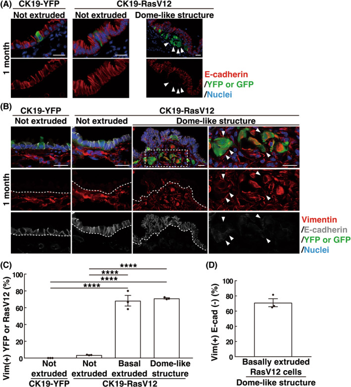
FIGURE 4.

EMT‐like features in basally extruded RasV12‐transformed cells in dome‐like structures. (A, B) Immunofluorescence images of E‐cadherin and vimentin in bronchial epithelia from CK19‐YFP or CK19‐RasV12 mice with tamoxifen treatment. Scale bars, 20 μm. (A) Arrowheads indicate E‐cadherin‐negative basally extruded RasV12 cells in the dome‐like structure. (B) Dashed lines delineate the basement membrane of the epithelial layer. The dotted area is shown at higher magnification in the righthand panels. Arrowheads indicate vimentin‐positive, E‐cadherin‐negative basally extruded RasV12 cells in the dome‐like structure. (C, D) Quantification of vimentin+ YFP or RasV12 cells (C) and vimentin+ E‐cadherin− basally extruded RasV12 cells (D). (C) ‘Not extruded’ encompasses both single and clustered nonextruded YFP or RasV12 cells; vimentin‐positive ratio of ‘not extruded’ was low, irrespective of the number of cells in cell clusters. Data are mean ± SEM n = 152 (not extruded in CK19‐YFP), 110 (not extruded in CK19‐RasV12), 57 (basal extruded in CK19‐RasV12), and 198 (dome‐like structure in CK19‐RasV12) cells from three mice. ****p < 0.001, one‐way anova with Tukey's test. (D) n = 124 cells from three mice.
