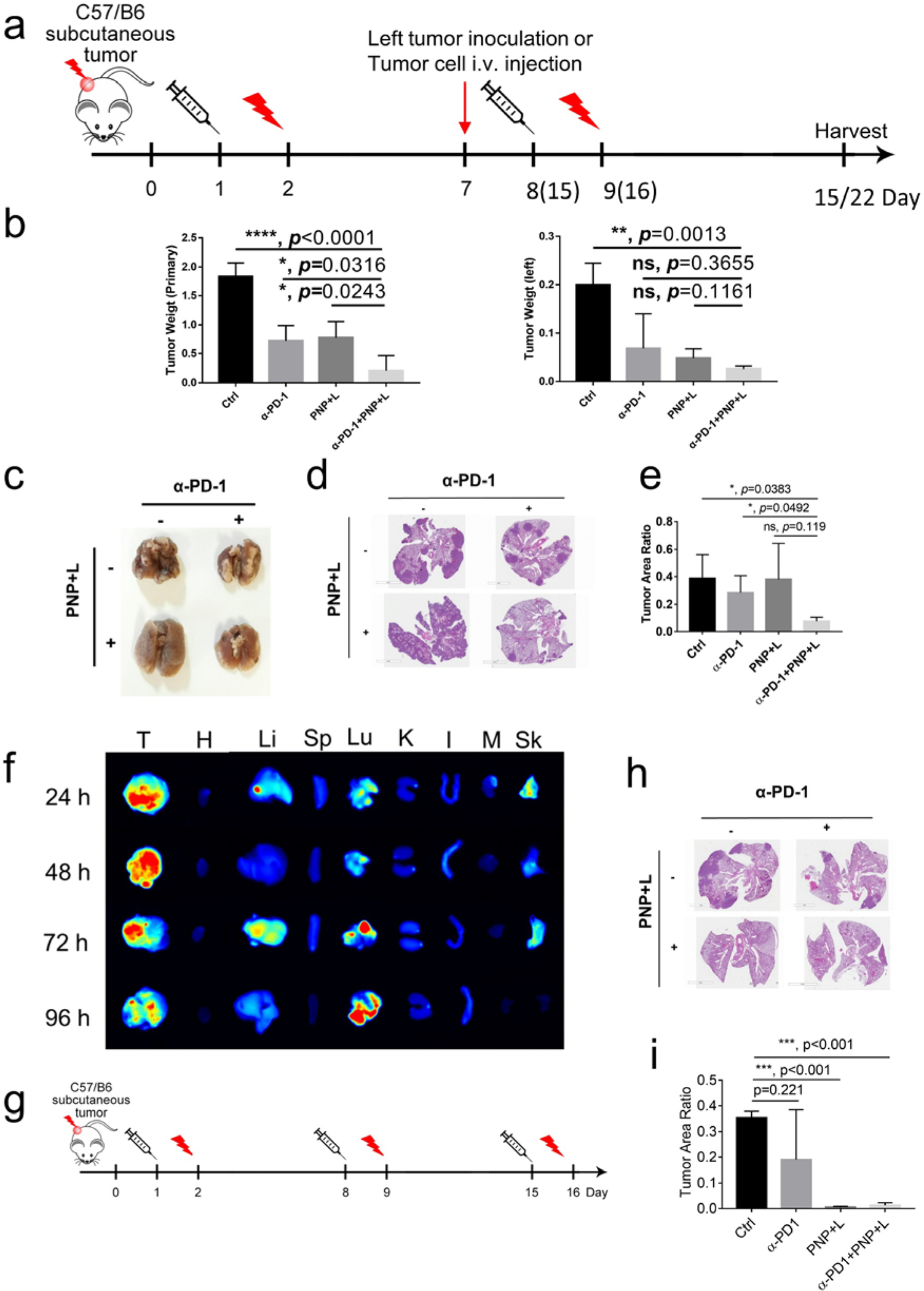
Figure 4: Synergistic PNP and immunotherapy for in vivo suppression of distant tumors and lung metastasis.

a) Experimental design to determine whether PDT and immunotherapy could prevent development of induced distant metastasis. MB49(1 × 10⁶ cells) tumor implants were first established at the right flank of C57BL/6 mice. When tumors reached the size of 100 mm3, mice received PBS or PNP (Day 1) followed by light treatment at the right flank 24 hours later (Day 2), and anti-PD-1 treatment was given twice a week. The same set of treatment was repeated once in the groups mimicking distant metastasis and twice in the groups mimicking lung metastasis. MB49 cells (1 × 10⁶ cells) were injected to the left flank on Day 7 to determine abscopal effects. To mimic lung metastasis, MB49 cells (1 × 10⁵ cells) were injected via tail vein on Day 7. b) The tumors on the right side were designated as primary tumors treated with light, and those on left side were designated as distant tumor (n=4). Tumors were harvested 15 days after the first treatment. Statistical analyses were performed with tumor weights on primary site and distant site. For the right (treated) site, tumor weight was 0.21±0.13 g in the combination treatment group, compared 1.83±0.12 g in the control group (p<0.0001), 0.72±0.13 g in the anti-PD-1 group (p=0.0316), and 0.78±0.14 g in the PNP+ PDT group (p=0.0243), respectively (left panel). The distant tumor in the combination group was also much smaller than that in the control (0.02 ± 0.001 gram versus 0.2 ± 0.02 gram, p=0.0013) even though not much smaller than the other two groups (right panel). c-e Lungs were harvested 22 days after the first treatment in the groups mimicking lung metastasis. c) Gross appearance of tumor nodules in the lungs. d) Hematoxylin and eosin staining of lungs. e) Statistical analysis of tumor area. The tumor/lung ratio in the combination group was smaller than the control (p=0.0383) and anti-PD-1 (p=0.0492) groups, but not the PNP plus light treatment group (p=0.119). f) fluorescent signal showing PNP accumulation in tumor and lung, suggesting development of spontaneous lung metastasis. (T: tumor, H: heart, Li: Liver, Sp: Spleen, Lu: Lung, K: Kidney, I: Intestine, M: Muscle, Sk: Skin). g) Schema showing the design to determine whether PDT and immunotherapy could prevent development of spontaneous lung metastasis. h) Hematoxylin and eosin staining of lungs. i) Statistical analysis of tumor area. Compared to the control, the PNP plus light treatment and the combination groups significantly decreased spontaneous lung metastasis (p<0.001).
