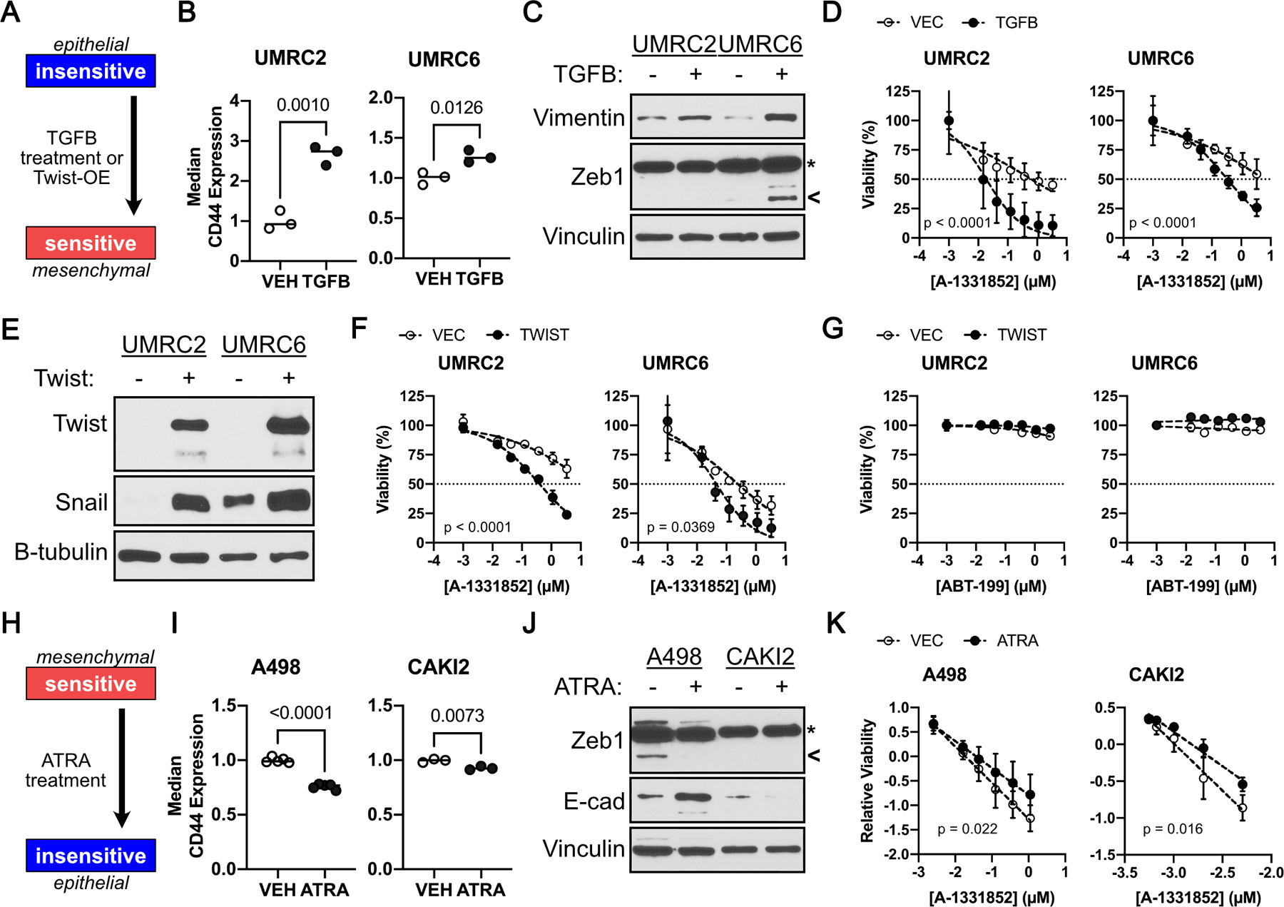
FIGURE 4.

A Mesenchymal Cell State Promotes BCL-XL Dependency. A, Schema showing experimental design to address sufficiency of mesenchymal state in promoting Bcl-xL dependency. CD44 levels, as determined by flow cytometry (B), and immunoblot analysis of the indicated proteins (C), in A-1331852 insensitive ccRCC cells that were treated with 10 ng/ml TGFβ for 3 days. D, Cell viability, as measured using CellTiter-Glo, in the indicated cells treated with A-1331852 for 7 days. Immunoblot analysis (E) and cell viability, as measured by Cell-TiterGlo, in the indicated insensitive ccRCC cells that were treated with the indicated concentration of A-1331852 (F) or ABT-199 (G) for 7 days. (H) Schema showing experimental design to address necessity of mesenchymal state in promoting BCL-XL dependency. CD44 levels, as determined by flow cytometry (I), and immunoblot analysis (J), in the indicated ccRCC cells that were treated with 1 µM ATRA for 3 days. K, Cell viability, relative to untreated DMSO controls, in the indicated cells treated with A-1331852 for 7 days. Data in (B) and (E) was normalized to CD44 levels in the untreated (Vehicle) control and was compared using the Student’s t-test (n ≥ 3, bar represents Mean, p-values are indicated). In (D), (F), (G), and (K), data were normalized for batch effects and compared using linear regression (n ≥ 3, mean±S.D., line represents best fit, p-values indicated are for difference in slopes i.e., interaction between A-1331852 and ATRA or TGFβ. In (D), (F), (G), and (K), all data points were plotted relative to the untreated DMSO controls in the given experimental arm, and all concentrations are presented as log10. In (C) and (J) arrowhead marks the ZEB1 band; whereas, asterisk marks a non-specific band, which doesn’t respond to cell state modifiers.
