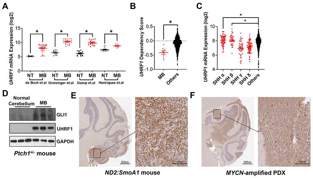
Figure 2. UHRF1 is overexpressed in SHH-MB.

(A) Meta-analysis comparing the expression of UHRF1 between normal brain tissue (NT) and patient derived MB tissue (MB) using the four indicated microarray datasets [42–45]. (B) The dependency on UHRF1 for patient-derived cancer cell line viability was compared between MB cell lines (MB) and non-MB cancer cell lines (Others) using the data from the DepMap project (Broad Institute). (C) Meta-analysis comparing the expression of UHRF1 among the four SHH subtypes (SHH α, β, γ and δ) and other subgroups of MB (Others), using microarray data from Cavalli et al [4]. (D) Normal mouse cerebella or SHH-MB tissue from Ptch1+/− mice were isolated and immunoblotted to determine the levels of GLI1, UHRF1 and GAPDH (n=3 mice per group). MB tissue from (E) ND2:SmoA1 or (F) patient-derived TRP53-mutant, MYCN-amplified MB xenograft (SJSHHMB-14-7196) mice were immunostained for UHRF1. Representative images are shown (n=3).
