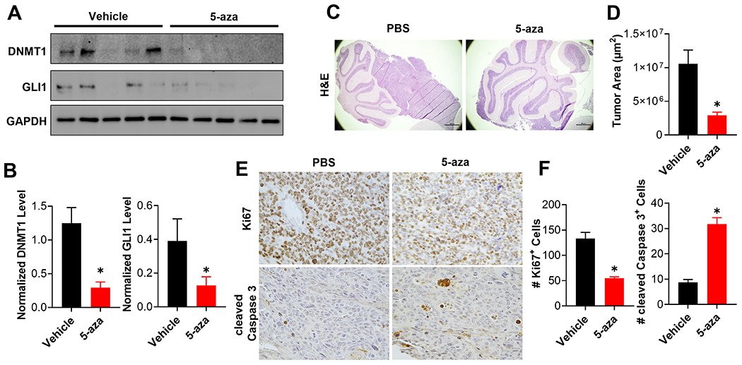
Figure 6. 5-aza attenuates SHH-MB progression in vivo.

(A) Murine MB cells, derived from a primary MB that had never been cultured ex vivo, were subcutaneously implanted into the flank of 10 CD1-Foxn1nu mice. When tumors reached ~ 100 mm3, these mice (n=5 mice per group) were treated with vehicle or 5-aza (5 mg/kg IV in PBS) every other day for up to 8 days. Residual tumor tissues were harvested 6 hours after the last injection, and subjected to immunoblotting of the resultant tumors for the indicated proteins (n=5 mice per group). (B) The level of DNMT1 (Left) or GLI1 (Right) was quantitated and normalized to that of GAPDH to calculate normalized protein level in each treatment group (n=5 mice per group). (C) Same MB cells as used in (A) were orthotopically implanted into the cerebella of CD1-Foxn1nu mice. These mice were treated 14 days after implantation with vehicle or 5-aza (5 mg/kg IV in PBS) every three days for 18 days. Representative H&E staining of the cerebella from these mice is shown (n=5 mice per group). (D) Tumor area was calculated from 4 independent mice per group and the results summarized in a bar graph. (E) Brain sections from the same mice were subjected to immunohistochemical staining for Ki67 or cleaved Caspase 3. Representative figures are shown (n=4 mice per group). (F) The number of Ki67 positive cells (Left) or cleaved Caspase 3 positive cells (Right) were quantitated in 4 random high magnification fields from each of the 4 mice in each group and the results summarized in bar graphs.
