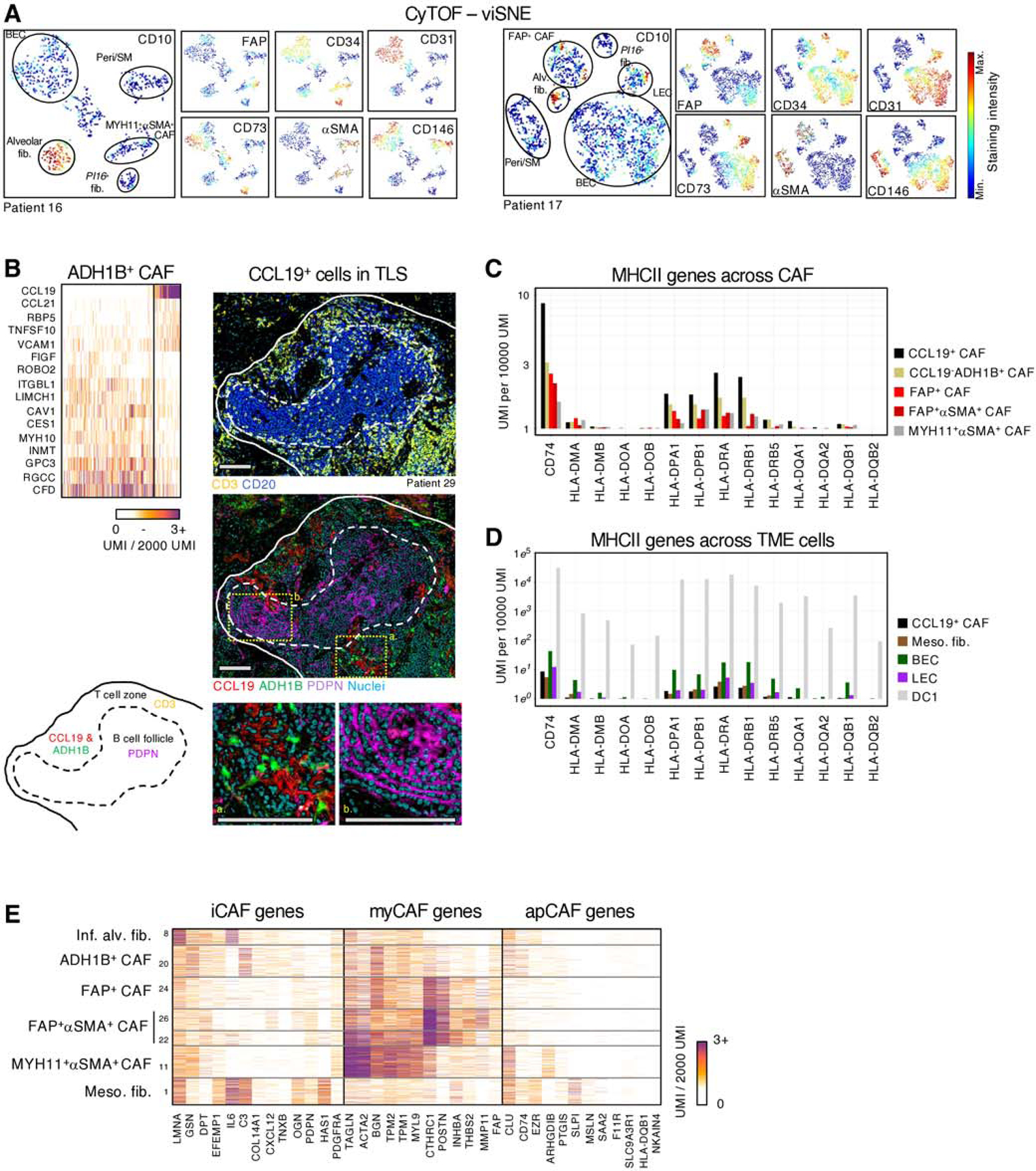
Fig. 2 |. Further characterization of CAF subsets in human NSCLC.

A, Stromal cell populations visualized with viSNE in CyTOF. EC, PvC and multiple fibroblast subsets can be distinguished with relatively few markers (CD10, CD31, CD34, CD73, FAP, CD146 and αSMA) B, (upper left panel) Highlighting CCL19 expressing cells within ADH1B+ CAF. These cells expressed high amounts of CCL19, CCL21, and VCAM1 and low levels of certain ADH1B+ CAF genes such as MYH10 and GPC3 (bottom and right panels). Multiplex IHC of a representative tertiary lymphoid structure. Podoplanin (PDPN) and CD20 marks follicular dendritic cells and B cells, respectively, in the B cell follicle, while the T cell zone is identified with CD3 staining. CCL19 and ADH1B staining show ADH1B+ fibroblasts surrounded by the secreted chemokine CCL19, specifically in the T cell zone. All scale bars are 100μm. C, Average expression of MHCII genes in each CAF subset. D, Average MHCII gene expression in classical antigen presenting cells, DC1, endothelial cells, and meso-like fibs. E, myCAF, iCAF and apCAF gene signatures (20,22) projected onto NSCLC CAF clusters.
