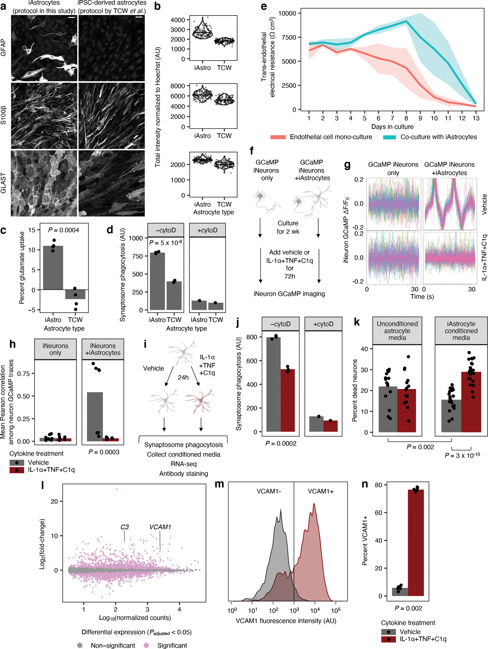
Fig. 1 |. iPSC-derived astrocytes (iAstrocytes) perform canonical astrocyte functions and recapitulate key aspects of inflammatory reactivity.

a, Representative immunofluorescence images of astrocyte markers in iAstrocytes (“iAstro”) vs. hiPSC-derived astrocytes generated using the protocol from TCW et al.16 (“TCW”). Scale bar: 60 μm. b, Quantification of data in a; data points represent fields of view collected over two replicates. c, Glutamate uptake (n = 4 wells). d, Phagocytosis of pHrodo-labeled rat synaptosomes (median pHrodo fluorescence by flow cytometry) with (n = 1 well) or without (n = 3 wells) cytochalasin D (cytoD). e, Barrier integrity of brain endothelial-like cells cultured alone (n = 6 wells) or with iAstrocytes (n = 5 wells); lines – group means, shaded bands around lines – 95% confidence intervals. f,g, Neuronal calcium activity traces of GCaMP iNeurons co-cultured with iAstrocytes treated with vehicle or IL-1α+TNF+C1q; traces from individual neurons are overlaid. h, Synchrony between neuronal calcium activity traces in iNeuron mono-cultures or iNeuron + iAstrocyte co-cultures treated with vehicle or IL-1α+TNF+C1q (n = 8 wells). i, Experiments assessing inflammatory reactivity. j, Phagocytosis of pHrodo-labeled rat synaptosomes by iAstrocytes treated with vehicle or IL-1α+TNF+C1q with (n = 1 well) or without (n = 3 wells) cytoD. k, Percentage of dead cells (TO-PRO-3 permeability) for iNeurons incubated with unconditioned astrocyte media with or without IL-1α+TNF+C1q (n = 14 or 16 wells, respectively) or astrocyte media conditioned by iAstrocytes treated with vehicle or IL-1α+TNF+C1q (n = 23 or 24 wells, respectively). l, Log-scaled fold change vs. average expression of differentially expressed genes (RNA-seq) induced by IL-1α+TNF+C1q in iAstrocytes (n = 3 wells). m, Representative histogram of cell-surface VCAM1 levels (flow cytometry) in iAstrocytes treated with vehicle or IL-1α+TNF+C1q. n, Percent of VCAM1+ iAstrocytes after treatment with vehicle or IL-1α+TNF+C1q (n = 4 wells). In panels c, h, k, and n, P values were calculated using the two-sided Mann-Whitney U test. In panels d and j, P values were calculated using the two-sided Student’s t-test. In panel l, P values were calculated and adjusted for multiple testing (Benjamini-Hochberg method) using DESeq2 (two-sided Wald test; see Methods).
