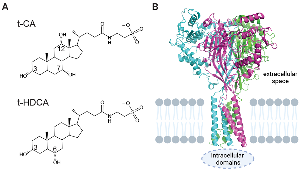
Figure 1. Bile acid and ENaC structures.

A, Structures of t-CA and t-HDCA, with positions of key functional groups numbered. Downward bonds face the hydrophilic side (α side) and upward bonds face the hydrophobic side (β side). Taurine conjugation adds a sulfonate group (pKa < 2), which remains ionized at physiologic pH. B, Cartoon of ENaC structure (pdb code 6BQN (Noreng et al., 2018)) colored by subunit with approximate location of membrane indicated. Created with BioRender.com
