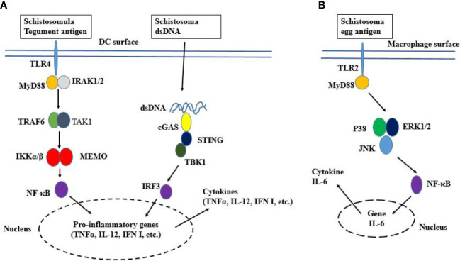
Figure 1.
Schematic representation of TLR signaling. (A) TLR4 localizes to the cell surface. It is activated by ligand binding, which leads to the dimerization of TLR and the recruitment of TLR domain-containing adaptor proteins. Then MyD88 activates IRAK which induces K63-linked polyubiquitination on TRAF6 itself and TAK1. The TAK1 activation leads to the activation of IKK complex NF-κB and cytokine genes transcription. The worm’s DNA is sensed by cGAS, resulting in the activation of STING-TBK1-IRF3 signaling and IFN-I response. (B) Egg activated TLR2 recruits MyD88, resulting in the phosphorylation of P38, ERK1/2, and JNK. Thereafter, NF-κB induces the expression of the IL-6 gene.

