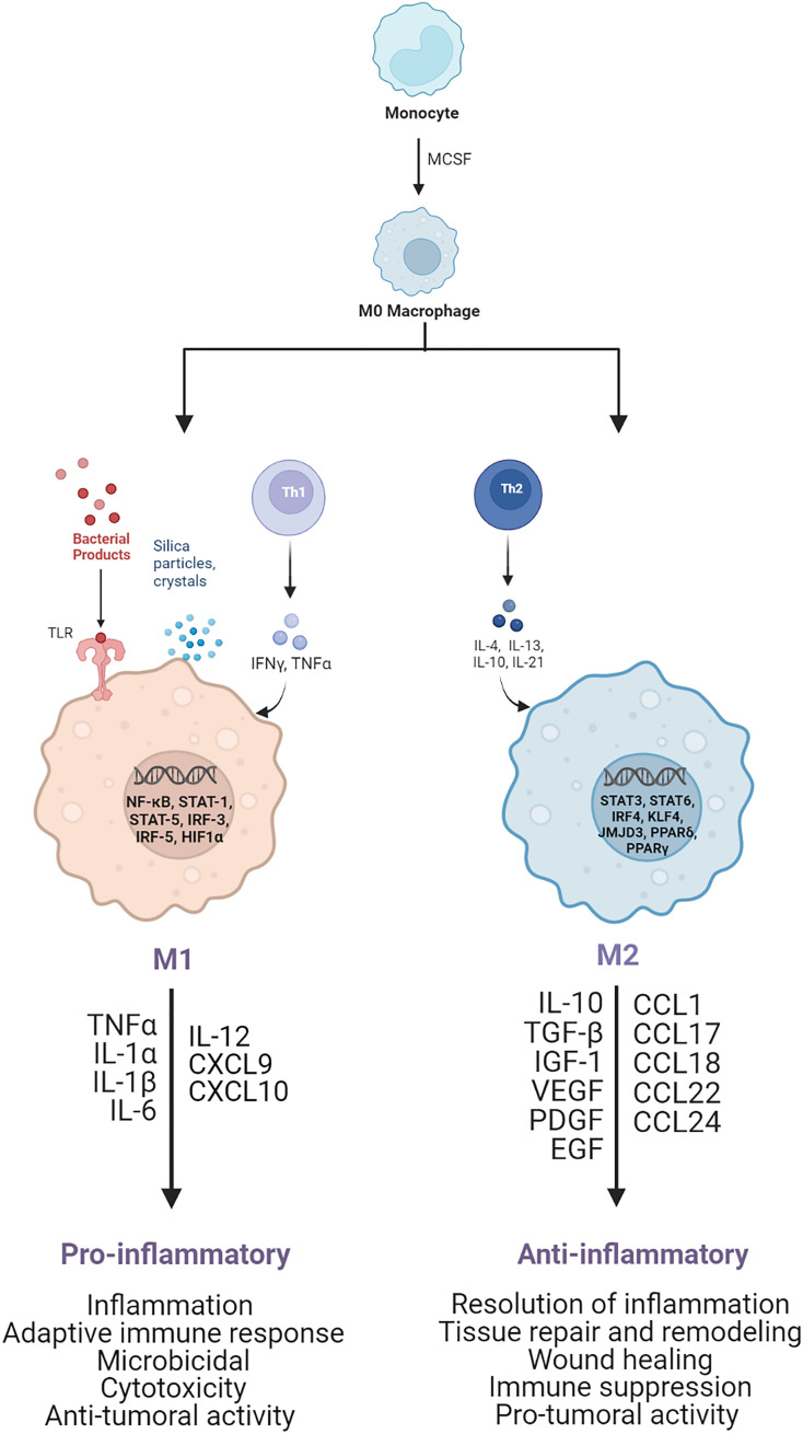
Figure 1.
Schematic of M1 and M2 macrophage activation. M1 pro-inflammatory macrophages are activated by TLRs ligands, such as LPS, or Th1 cytokines, such as TNF-α and IFN-γ. After activation, several transcription factors are involved, such as NF-kB, STAT1, STAT5, IRF3, and IRF5, leading to the release of pro-inflammatory cytokines and chemoxines, including TNF-α, IL-1α, IL-1β, IL-6, IL-12, CXCL9, and CXCL10, which exert microbicidal and anti-tumoral functions. M2 anti-inflammatory macrophages are polarized by Th2 cytokines, such as IL-4 and IL-13, which activate transcription factors, including STAT3, STAT6, IRF4, KLF4, JMJD3, PPARδ, PPARγ. As result the M2 activated macrophages release anti-inflammatory cytokines and chemoxines including IL-10, TGF-β, CCL17, CCL18, and CCL22, which promote wound healing, tissue repair and regeneration, immune-suppression and tumor grow and diffusion.

