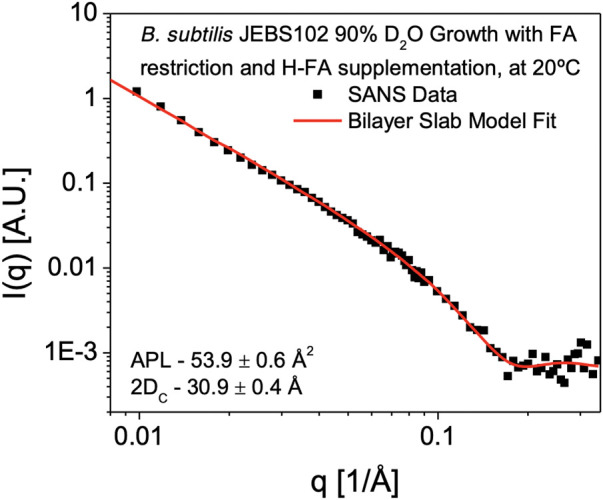
FIGURE 4.

SANS Measurement of Cell Membrane hydrophobic Thickness. SANS data obtained from B. subtilis JEBS102 cells grown in 90% D2O media, induced to block expression of fabF and rescued with supplementary FAs, H-anteiso-pentadecanoic acid (a15:0) and H-normal-hexadecanoic acid (n16:0) in 85% D2O resuspension media is plotted as scattered intensity, I(q), as a function of scattering wavevector, q (Å−1). Error bars correspond to ± σ. The experimental data are shown superimposed with the best-fit (red solid line) using a self-consistent lamellar form factor (Tan et al., 2021) and the parameters described in Table 1, 2 revealing an average hydrophobic thickness of 30.9 ± 0.4 Å. The form factor is consistent with contrast being introduced exclusively within the hydrophobic portion of the cell membrane.
