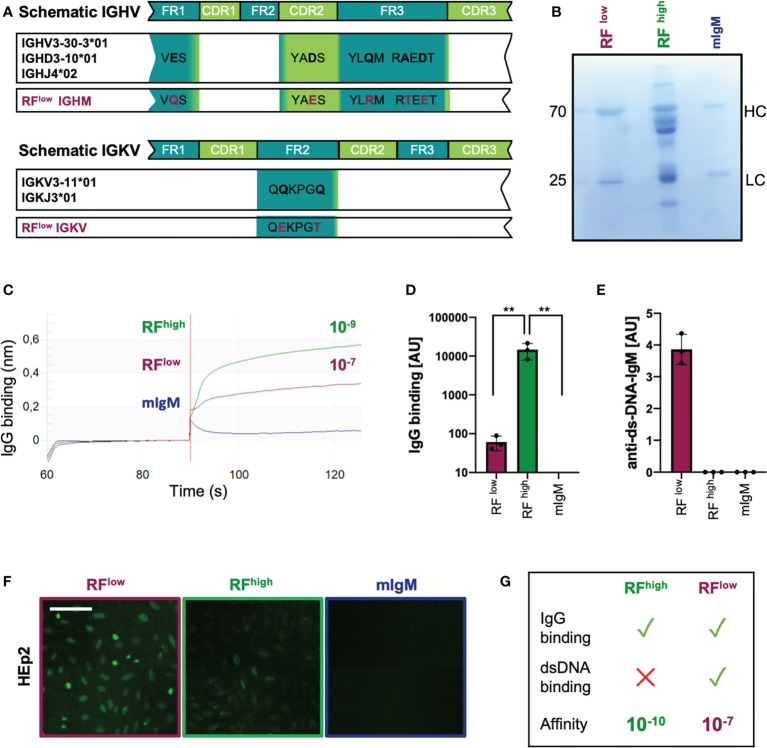
Figure 3.
Recombinant low-affinity RF is polyreactive and binds DNA. (A) Schematic representation of the immunoglobulin heavy and light variable genes (IGHV and IGLV, respectively) of the recombinant purified low-affinity RF as compared to the closest germline respective alleles. Mutations are highlighted in purple. IGHM, immunoglobulin heavy constant mu; IGVK, immunoglobulin variable kappa. (B) Coomassie blue-stained SDS-PAGE showing recombinant monoclonal (in-house purified) low affinity RF (RFlow), commercial RF from Rheumatoid Arthritis patients (RFhigh) and monoclonal control IgM (mIgM) under reducing conditions (with β-mercaptoethanol). The image is representative of three independent experiments. (C) IgG-binding affinity of RFlow (purple line), RFhigh (green line) and monoclonal IgM (blue line) measured by bio-layer interferometry. KD (dissociation constant) was calculated by the software. The experiment shown is representative of 3 independent experiments. (D) Anti-IgG IgM concentrations detected in purified RFlow (purple bar, n = 3), RFhigh (green bar, n = 3) and mIgM control (blue bar, n=3) measured by ELISA (coating: human IgG). Mean ± SD, statistical significance was calculated using ordinary one-way ANOVA with Tukey’s multiple comparisons test. **p < 0,01. (E) Anti-dsDNA-IgM concentrations of RFlow (purple bar, n = 3), RFhigh (green bar, n = 3) and IgM control (blue bar, n = 3) measured by ELISA (coating: calf-thymus dsDNA). Mean ± SD. Results are representative of three independent measurements. (F) HEp-2 slides showing anti-nuclear structure-reactive IgM (ANA). Purple square: RFlow; green: RFhigh; blue: mIgM control, respectively. Scale bar 65 µm. Green fluorescence indicates IgM binding to HEp-2 cells. Images are representative of three independent experiments. (G) Schematic summary of the characteristics of RFhigh and RFlow.

