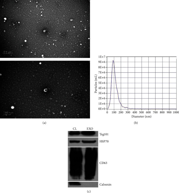
Figure 1.

Characterization of exosomes. Transmission electron microscopy images of exosomes isolated from plasma (a). The size distribution of exosomes indicated by nanoparticle tracking analysis (b). Western blot analysis of Alix, Tsg101, CD63, and Calnexin in plasma-derived exosomes (c).
