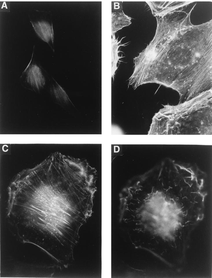
FIG. 2.
Actin staining of CNF1-treated HeLa cells. HeLa cells were treated with GST-CNF1 (500 ng/ml) for 0 (A), 6 (B), and 24 (C and D) h. Thereafter, the cells were fixed and the F-actin was stained with rhodamine-phalloidine. (C and D) Micrographs from the same cell, with focusing below the apical cell surface (C) and on the apical surface of the cell (D). For all panels, a 100× oil immersion objective was used.

