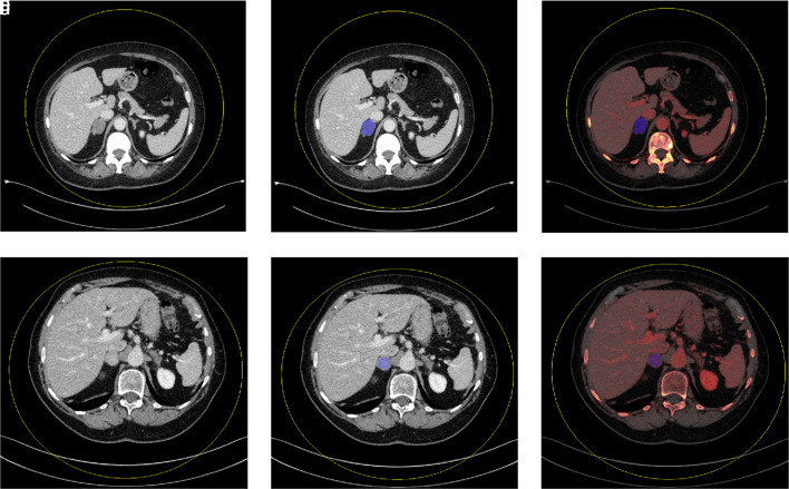
Figure 3.
a-f. Mixed density images demonstrate mixed density axial images of single-slice adrenal segmentation in a 61-year-old woman with an adrenal adenoma (a) and an 83-year-old woman with an adrenal metastasis (d). Mixed and iodine material density images demonstrate single-slice adrenal segmentation of an adenoma (b-c) and a metastasis (e-f).

