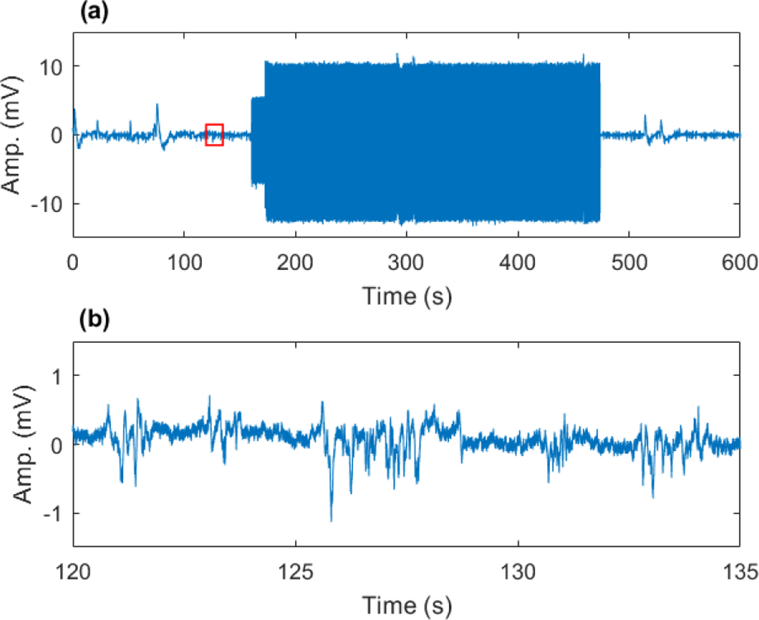
Fig. 1.

Example of interferences in extracellular neural recording caused by fMRI scanning at UHF. Scanning artifacts are tens of millivolts (a) compared to extracellular field potentials, which are hundreds of microvolts (b). Subplot (b) corresponds to small (red) boxed region in (a) in the absence of fMRI scanning (baseline period).
