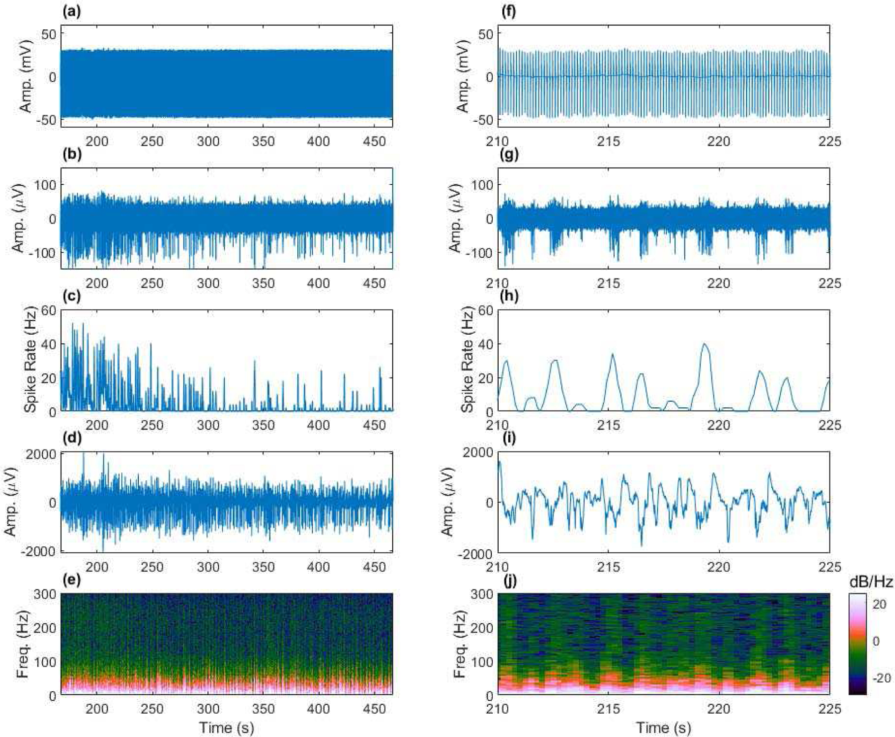
Fig. 8.

Removing fMRI artifacts from spontaneous EFPs (1–6000 Hz) acquired from the anesthetized rat cortex to achieve fully sampled neural recording during fMRI. (a) Artifact contaminated data, (b) artifact removed EAPs (300–6000 Hz), (c) spike rate computed from (b), (d) artifact removed LFP (1–300 Hz), and (e) multi-tapered spectral analysis of (d). Shorter time scale views of (a)-(e) provided in (f)-(j). Example from Rat C3, channel 9 during an increase in isoflurane anesthesia concentration leading to reduced neural activity.
