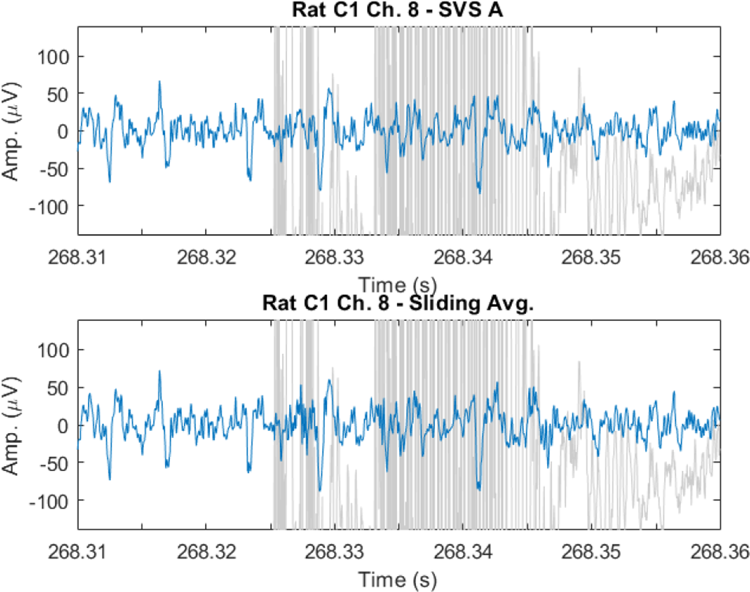
Fig. 9.

EAPs recovered from spontaneous EFP recorded during fMRI artifacts in the anesthetized rat. EAP signal shown in blue over artifact contaminated data in light gray. EAPs are observable in artifact denoised data using both SVS method A (top) and the sliding template subtraction approach (bottom).
