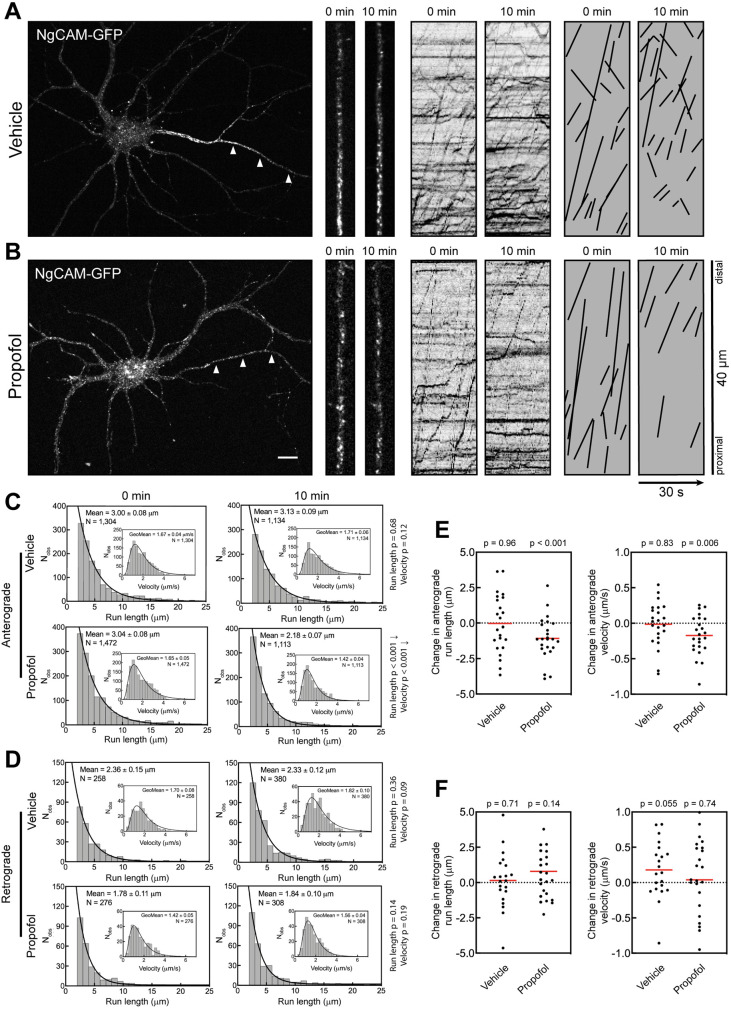
FIGURE 3:
Propofol attenuates axonal transport of the KIF5C cargo NgCAM. (A, B) Representative images of 7–8 DIV hippocampal neurons expressing NgCAM-GFP. Arrowheads indicate the axon. High-magnification images and kymographs show vesicles and axonal transport of NgCAM-GFP vesicles before and 10 min after treatment with (A) vehicle (DMSO) or (B) 10 µM propofol. For clarity, transport events from kymographs are redrawn as black lines. Scale bar: 10 µm. (C, D) Histograms of axonal run lengths and velocities for axonal vesicles visualized with NgCAM-GFP. (E, F) Plots of the change in run length and velocity for each neuron from 0 to 10 min treatment with vehicle (DMSO) or propofol. DMSO: 21 cells; propofol: 23 cells.

