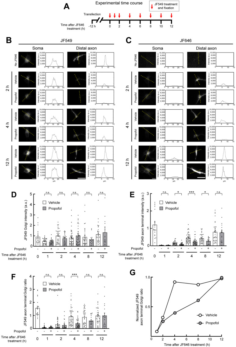
FIGURE 4:
Propofol delays delivery of membrane proteins to the distal axon. (A) Schematic showing the experimental design. Twelve hours after transfection with NgCAM-Halo, neurons were incubated in JF646 dye to saturate existing NgCAM-Halo binding sites. After JF646 washout, NgCAM-Halo was treated with JF549 and fixed at the indicated time points. (B, C) Representative images of NgCAM-Halo labeled by JF549 (B) or JF646 (C) in the soma and distal axon of neurons, visualized and fixed at the indicated time points after JF646 saturation. Dotted lines indicate the areas from which intensity plot profiles were generated. Scale bars: 10 µm. (D, E) Quantification of JF549-labeled NgCAM-Halo at the Golgi (D) and the distal axon (E). (F) Quantification of the axon-to-Golgi JF549 ratio. (G) Golgi-to-axon ratio values plotted as a fraction of the maximal 12 h time point. P values: * < 0.05; *** < 0.001.

