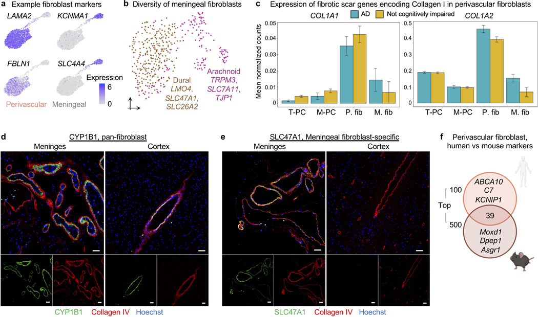
Extended Data Fig. 7. Specialization and functions of human brain fibroblasts.
a, Expression of example markers demarcating perivascular from meningeal fibroblasts.
b, UMAP of 428 meningeal fibroblast nuclei, subclustering into anatomically segregated dural and arachnoid space fibroblasts.
c, Expression of the genes constituting the major fibrotic scar component Collagen I in pericytes and fibroblasts. Collagen I is composed of two components, COL1A1 and COL1A2. Column annotations: T-PC = solute transport pericyte and M-PC = Extracellular matrix regulating pericyte, P. FB = Perivascular fibroblast, and M. FB = Meningeal fibroblast.
d-e, Protein immunostaining validation of polarized expression of human brain meningeal and perivascular fibroblast pumps: the common marker CYP1B1 (a, serves as a control) and the meningeal fibroblast-specific influx pump SLC47A1 (b). Scale = 50 μM.
f, Overlap between the top 100 perivascular fibroblast-like cell markers and those identified in mice. A more lenient set of 500 (instead of 100) mouse markers52 were used for comparison to ensure claims of species-specificity were robust. Note: the species-conservation of a cell type marker depends on speciesspecific changes in the given cell type and changes amongst the remaining background cell types.

