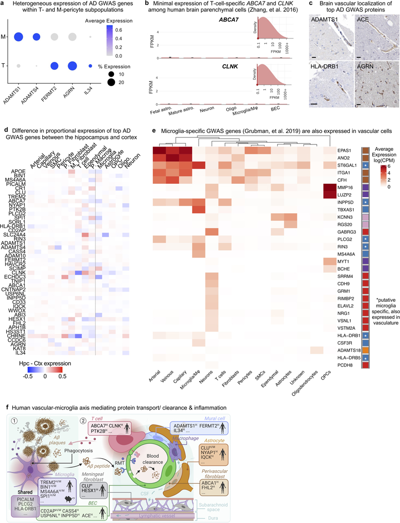
Extended Data Fig. 9. Re-evaluation and characterization of top AD GWAS genes expressed in the human brain vasculature.
a, Heterogeneous expression of AD GWAS genes across T- and M-pericyte subtypes.
b, RNA-seq data of the predicted T cell-specific AD GWAS genes EPHA1 and ABCA7 in an independent dataset81, corroborating minimal expression across resident/ parenchymal brain cells.
c, Immunohistochemical confirmation of vascular localization of proteins encoded by 12 top AD GWAS genes from (a). Scale bar = 25 microns. Arrowheads in APOE point to signal around larger diameter vessels, consistent with predicted SMC expression. Image credit: Human Protein Atlas (http://www.proteinatlas.org)25,75.
d, Heatmap comparing expression patterns of top AD GWAS genes in the hippocampus and superior frontal cortex: e.g., several microglia-expressed GWAS genes like APOE, MS4A4A, and TREM2 are more highly expressed in hippocampal compared to cortical microglia/ macrophages.
e, GWAS genes found to be expressed specifically in microglia among cells captured using the conventional nuclei isolation process (from Grubman, Chew, Ouyang, et al. 201917) are also expressed in vascular cells (asterisks).
f, Summary of AD GWAS genes enriched in microglia and vascular cells mediating common pathways in protein clearance and inflammation. Mouse and human superscripts denote whether expression has been confirmed in that species for a given gene. Proposed model is described in Discussion.

