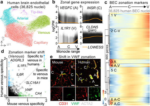
Figure 2. Organizing principles of human brain endothelial cells.
a, UMAP of 36,825 human brain endothelial cell (BEC) nuclei, colored by zonation.
b, Zonal expression of transcripts across human BECs ordered by Monocle pseudotime. LOWESS regression line (orange) and density of black lines (counts) correspond with expression levels. A = arterial, C = capillary, and V = venous.
c, Heatmap of zonation-dependent gene expression in human BECs.
d, Scatter plot depicting the specificity of transcripts for venous BECs in mice12 versus humans. Venous specificity score = avg(logFC(vein/cap), logFC(vein/art)). For example, VWF is predicted to be more specific to venous BECs in mice than it is in humans. See Extended Data Fig. 9 for arterial and capillary specificity plots.
e, Immunohistochemical validation of VWF specificity to venous BECs in mice but not in humans. Scale bar = 50 microns.

