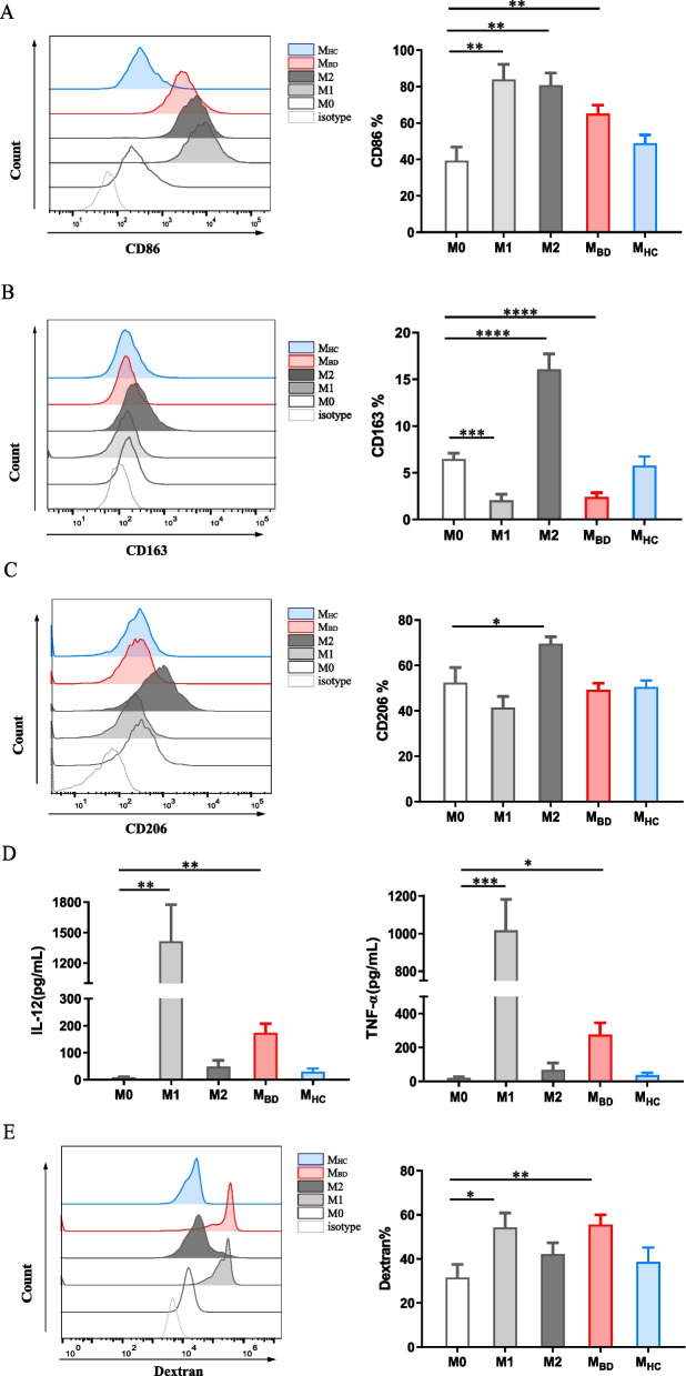
Fig. 1.
BD serum promotes M1-like macrophage polarization. Resting macrophages (M0) were stimulated with M1 condition (100ng/ml LPS+ 20ng/ml IFNγ), M2 condition (20ng/ml IL-4+ 20ng/ml IL-13), BD serum or HC serum for 48 h. A–C Representative histograms (left) and summary (right) of CD86, CD163 and CD206 expression level of macrophages stimulated with M0 (n=6), M1 (n=6), and M2 (n=6) conditions, as well as BD (n=12) serum and HC (n=12) serum. Data were expressed as mean±SD and were analyzed using one-way ANOVA. D IL-12 and TNF-α production by macrophages stimulated with M0 (n=6), M1 (n=6), and M2 (n=6) conditions, as well as BD (n=12) serum and HC (n=12) serum. Data were expressed as mean±SD and were analyzed using Kruskal-Wallis test. E Representative histograms (left) and summary (right) of dextran uptake by macrophages stimulated with M0 (n=7), M1 (n=7), M2 (n=7) conditions, and BD (n=9) serum and HC (n=9) serum. Data were expressed as mean±SD and were analyzed using one-way ANOVA. *, p<0.05; **, p<0.01; ***, p<0.001, ****, p<0.001. MBD, BD serum-treated macrophages; MHC, HC serum-treated macrophages

