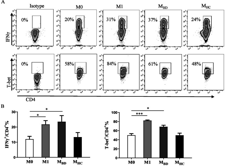
Fig. 2.
BD serum-treated macrophages facilitate Th1 differentiation. Naive CD4+ T cells were incubated with M0, M1, M2, BD serum- and HC serum-treated macrophages in Th1 condition (5μg/ml anti-CD3, 5μg/ml anti-CD28, 5μg/ml anti-IL-4, and 10ng/ml IL-2) for 5 days. A Representative flow cytometry plots and B summary of IFNγ and T-bet [n(M0)=3, n(M1)=3, n(M2)=3, n(MBD)=5, n(MHC)=5] expression levels in CD4+ T cells. Data were shown as mean±SD. *, p<0.05; **, p<0.01; ***, p<0.001, ****, p<0.001 by one-way ANOVA. MBD, BD serum- treated macrophages; MHC, HC serum- treated macrophages

